Central Line Placement Internal Jugular Procguide Internal Jugular Central Line
The syringe was removed and a guidewire was advanced into the introducer needle.
Central line placement internal jugular. The risk of complications of central line placement varies with the experience of the operator and the conditions emergency vs. Optimal positioning of right sided internal jugular venous. Central venous catheterization intra atrial electrocardiography peres height formula. Patient preparation and position for right ij central line placement.
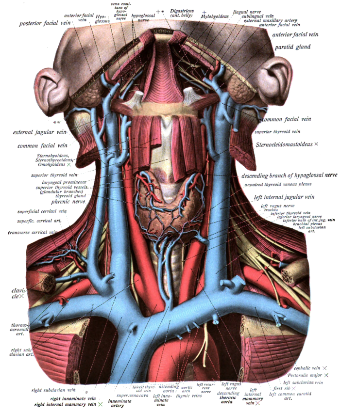
Venous blood was withdrawn. Using real time out of plane guidance the introducer needle was inserted into the internal jugular vein under direct ultrasound visualization. The internal jugular vein was identified using the ultrasound. Central venous line placement is typically performed at four sites in the body.
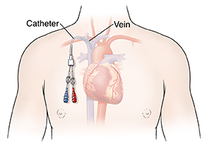
Hover onoff image to showhide findings. A central venous catheter cvc also known as a central line central venous line or central venous access catheter is a catheter placed into a large veinit is a form of venous accessplacement of larger catheters in more centrally located veins is often needed in critically ill patients or in those requiring prolonged intravenous therapies for more reliable vascular access. It can determine the exact position intraoperatively and can justify a delayed postoperative chest x ray to confirm cvc line tip placement. A long catheter may be advanced into the central circulation from the antecubital veins as well.
The right or left internal jugular vein ijv or the right or left subclavian vein scv. Shows compression of left internal jugular vessel. Still image of a peripheral vein. Alternatives include the external jugular and femoral veins.
Click image to align with top of page. The guidewire was. Central venous catheters cvc are routinely placed in patients undergoing major. Tap onoff image to showhide findings.
Anesthesia was achieved over the vein using 1 lidocaine. Risks associated with central venous catheterization include infectious mechanical and thrombotic complications. Still image of internal jugular vein in transverse view. This video demonstrates the technique for placement of a central venous line in the internal jugular vein and considers complications and how to avoid them.
Linear probe with sterile sheath cover. Nonetheless some general statements can be made and used when obtaining consent from a patient.


